Pneumothorax means air within the space between the lung and the chest wall called the pleura. It is commonly diagnosed on chest X-ray.
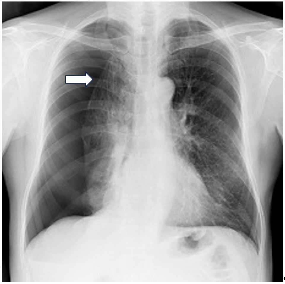
Right pneumothorax – see arrow for the edge of the collapsed lung, image courtesy of MedlinePlus from the National Library of Medicine, USA.
This can happen for several reasons, such as: •Trauma •Medical procedures or surgery •Primary spontaneous pneumothorax in young people with otherwise normal lungs •Secondary pneumothorax due to underlying lung disease
In most cases, a small pneumothorax can be watched by X-ray and may not need treatment. Larger ones may need a needle or chest tube / drain inserted to let out the air until the lung heals.
Primary Spontaneous Pneumothorax
This usually occurs in young slim adults, more commonly men than women, when a weak area of lung called a bleb or bulla bursts. The rest of the lung is normal.
If the pneumothorax is small, it may be watched without treatment. Larger ones will need to be treated with aspiration by a needle or with a chest drain. If the lung fails to re-expand, if it happens for a second time or if the person does scuba diving or is aircrew, then surgery will be recommended to prevent further episodes of pneumothorax.
Surgery can usually be performed keyhole (VATS or thoracoscopically) and involves removing the blebs or bullae along with making the lung stick to the chest wall to remove the space between them. This can be done by pleurectomy – removing the lining layer, pleural abrasion – by scraping with a rough pad, or by sterile talc, all aiming to make a strong scar between the lung and the chest wall to prevent further collapse.
Secondary Pneumothorax
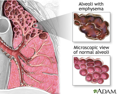
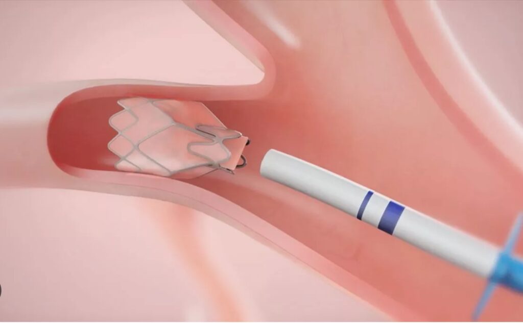
This occurs in patients with underlying significant lung disease such as emphysema, COPD, interstitial lung disease. Treatment is not as straightforward as above as most of the lung is affected and will usually require hospital admission. Treatment may be by chest drain and pleurodesis, surgery, or by placing endobronchial valves to block air to the affected part of the lung that is the cause of the leak.
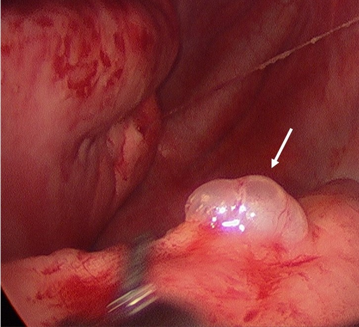
Image courtesy of Medline Plus from National Library of Medicine, USA