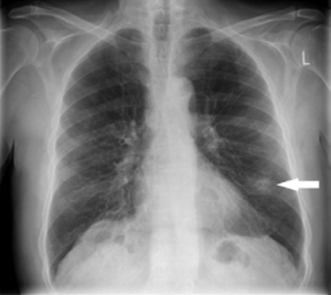
Lung Nodules

Picture from the American Thoracic Society,
“What is a lung nodule” patient information series.
https://www.thoracic.org/patients/patient-resources/resources/lung-nodules-online.pdf
Lung Nodules
Lung nodules are small volumes that are more solid than the normal surrounding lung tissue. They are commonly found on chest CT or MRI scans taken for screening or for other reasons such as a chest infection. By far the majority (90%+) of these are benign and not a significant concern. Most are scars from old infection or inflammation and can be left alone, others are difficult to tell whether they are benign or an early cancer. Various online calculators will predict the percentage risk whether the nodule is a cancer for a particular patient, based on their age, sex, smoking and family history along with the way the nodule looks on the scan.
Interval / Follow-up Scans
Small (up to 10mm / 1cm) nodules that do not look worrying will often be “observed” by follow up CT scans after an interval of 3 – 12 months and checking whether they grow. Nodules that do not grow over a period of two years are very unlikely to be a cancer. Either a biopsy or removal is generally recommended where the nodule is seen to grow.
PET-CT Scans
A PET-CT scan is often helpful in showing whether the nodule is a concern. A “hot” nodule lights up on the scan as it is using energy, if it is “cold” it is much more likely to be benign. However, both infection and cancer can show up as “hot” on the PET-CT scan and the scan can miss some cancers as “cold” nodules.
Biopsy
Larger nodules that are relatively close to the outside surface of the lung may be biopsied by a radiologist with a CT scan (CT biopsy), those closer to the centre and nearer the airways (bronchus) may be biopsied by navigation bronchoscopy or radial EBUS. However, sometimes the only way to find out is to remove them with surgery.
Surgery for Lung Nodules
Removing lung nodules is a major part of the work of the Thoracic Surgeon. Nodules that are known to be benign are generally only removed if they are causing problems such as persisting infection or blockage of an airway. However, removal is required if there is any doubt or may be required as treatment for a cancer.
For diagnosis, peripheral nodules (meaning close to the outside lung surface) can usually be removed with a simpler operation such as a wedge resection. If the nodule is known or found to be lung cancer, then a larger part of the lung is usually removed for potential cure – a segmentectomy or lobectomy.
In most cases, the surgery can safely be performed via keyhole or minimally invasive or thoracoscopic or VAT techniques – all these terms mean the same.
At King’s College Hospital, Dubai Hills, Dr. James Douglas Aitchison offers minimally invasive (VAT) surgery for diagnosis of lung nodules and for surgical treatment of lung cancers where appropriate. He has performed several hundred biopsies and several hundred thoracoscopic lobectomies for lung cancer.
Lung Nodule Diagnoses
Benign Causes
Infection
Granulomatas
Histoplasmosis
TB (Tuberculosis)
Atypical mycobacterium
Other Infections
Bacterial Abscess
Echinococcus
Pneumocystis
Aspergillus
Benign Neoplasms
Hamartoma
Lipoma
Neurofibroma
Leiomyoma
Angioma
Vascular
AV (arteriovenous) malformation
Pulmonary varix
Haematoma
Pulmonary infarct
Developmental
Bronchogenic cyst
Inflammatory
Rheumatoid nodule
Wegener’s granulomatosis
Sarcoidosis
Others
Rounded atelectasis
Lymph nodes
Mucocele
Cancers
Lung Origin
Adenocarcinoma
Squamous cell carcinoma
Small cell carcinoma
Spindle cell tumour
Carcinoid tumour
Metastatic (Other cancer spread)
Breast
Colon
Kidney
Sarcoma
Head and neck
Melanoma
Germ cell
Others
Lymphoma
Plasmacytoma
Schwannoma
